|
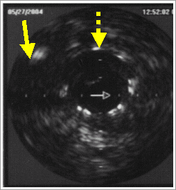
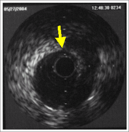
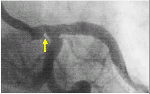
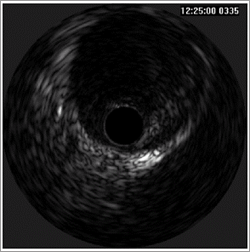
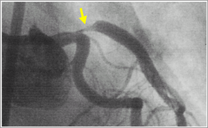
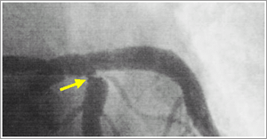
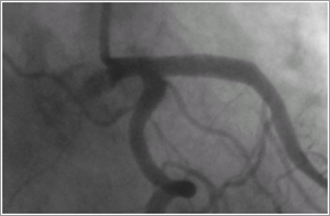
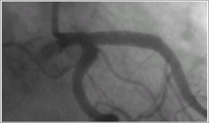
A 63-year-old male was referred for PCI for the ostial stenosis of left anterior descending coronary artery (LAD). He had complained of exertional angina (Canadian Society angina classification type III). He was a 60-pack year current smoker. His electrocardiogram showed T wave inversion on anterior chest leads and his left ventricular ejection fraction on echocardiography was 71%. The level of troponin-I was slightly elevated and other cardiac enzymes were normal. A diagnostic coronary angiogram revealed critical, eccentric stenosis in the ostium of LAD (Fig. 1) and intravascular ultrasound (IVUS) showed large amount of soft plaque in the ostium of LAD. After predilation using 3.5*20mm balloon, 3.5*18mm Cypher® stent was deployed for the ostium of LAD. However, after successful deployment of stent for the ostium of LAD, significant narrowing was developed in the ostium of LCX (Fig. 2). We thought that this narrowing of the ostium of LCX was caused by plaque shifting from the ostium of LAD. So, we performed IVUS for LCX. The IVUS for LCX showed very minimal soft plaque in ostium of LCX (Fig. 2), suggesting significant narrowing of the ostium of LCX was developed by spasm. After then, we performed another angiogram. The angiogram showed significant narrowing in d-LM (Fig 3). But, IVUS for LM showed minimal soft plaque in distal portion (Fig 3). This narrowing of d-LM was also developed by spasm. After intracoronary injection of nitroglycerin, these vasospasms of the ostium of LCX and d-LM were relieved (Fig 4). On follow-up coronary angiogram performed one week after stenting, the stent of LAD was patent and narrowings of the ostium of LCX and d-LM were not observed (Fig 5). No changes of images of the ostium of LCX and d-LM on follow-up IVUS.
|
Fig. 1. A diagnostic coronary angiogram revealed critical stenosis in the ostium of left anterior descending artery. The ostium of left circumflex artery and distal left main artery appeared normal.
 |
| |
Fig. 2. After stenting for the ostium of left anterior descending artery (LAD), severe narrowing was developed in the ostium of left circumflex artery (LCX). But, intravascular ultrasound showed very minimal plaque in the ostium of LCX. Lined arrow indicates the ostium of LCX and broken arrow indicates the ostium of LAD.
|
| |
Fig. 3. After stenting for the ostium of left anterior descending artery, narrowing was developed in distal left main artery (d-LM). Intravascular ultrasound showed minimal plaque in d-LM. Lined arrow indicates spasm in d-LM.
|
| |
Fig. 4. After intracoronary injection of nitroglycerin, vasospasms of the ostium of left circumflex and distal left main artery were relieved.
 |
| |
Fig. 5. On follow-up coronary angiogram performed one week later after stenting, the stent of left anterior descending artery was patent and narrowings of the ostium of left circumflex and distal left main artery were not observed.
 |
|


