이달의 증례
| Defining Coronary Artery Perforation with Ultrasound Contrast Agent | |||
|---|---|---|---|
| 년도 | 2018년 11월 | ||
| 카테고리 | 이달의 kcj Hot Article | ||
| 저자 | Young Jin Youn , MD, PhD1, Salman Khalid , MD2, Michael Azrin , MD, PhD2, and Juyong Lee , MD, PhD2 | ||
| 소속 | 1 Division of Cardiology, Department of Internal Medicine, Wonju College of Medicine, Yonsei University, Wonju, Korea. 2 Division of Cardiovascular Medicine, Calhoun Cardiovascular Center, University of Connecticut School of Medicine, Farmington, CT, USA. | ||
| 첨부파일 1 | 201811_01.jpg | ||
- 관리자
- 등록일 : 2018.10.24
- Hit 1,241
After recanalization and stenting a chronic total occlusion of the left anterior descending coronary artery (LAD), coronary artery perforation (CAP) was detected in the LAD (Figure 1A and Supplementary Video 1), but it was not definitive whether this CAP emptied into the pericardium (Ellis classification III) or into the right ventricle (RV) (Ellis classification III cavity spilling).1) Bedside echocardiography showed no definite pericardial effusion. Despite prolonged balloon inflation, spontaneous sealing was not achieved. To define the direction of the CAP, 2 mL of ultrasound contrast agent (UCA), Definity (Lantheus Medical Imaging, North Billerica, MA, USA) was injected into the LAD. The rapid and dense appearance of UCA in the RV on echocardiography confirmed that the CAP was connected to the RV (Figure 1B and Supplementary Video 2). Because there was felt to be no risk of cardiac tamponade the procedure was completed. The patient was hemodynamically stable at discharge and there were no subsequent complications at 1-month follow-up. Conservative management for this type of CAP is debated, but only a medium or large sized fistula is considered as a cause of heart failure and myocardial ischemia.2)
원문보기: https://e-kcj.org/DOIx.php?id=10.4070/kcj.2018.0179
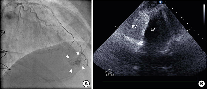 |
Figure 1. Coronary angiography and contrast echocardiography. (A) Post-stenting angiography shows CAP in the LAD (white arrowheads), but it is not definitive whether this perforation emptied into the pericardium (Ellis classification III) or into the RV (Ellis classification III cavity spilling). (B) After injection of UCA into the LAD, the rapid and dense appearance of UCA in the RV on echocardiography confirms the CAP of Ellis classification III cavity spilling. |
댓글 0
댓글 쓰기
[댓글 목록]
- 학회소개
- 학술 행사
- 학회지(KCJ)
- 심장학
최신지견 따라잡기
- 진료지침
- 자료실
- 회원공간
- 공지사항